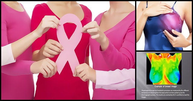
Through early detection, one can prevent the development of life-threatening breast cancer. When detected smaller or nonpalpable, breast cancers are more treatable, thereby associated with a more favorable prognosis.
Breast cancer screening identifies breast cancer in women who have no physical symptoms. At present, there is a debate about which methods should be used for screening, and how often women should be screened. Of course, everyone wants to choose the safest method.
In 2012 alone, the number of patients diagnosed with breast cancer reached up to 1,617,149 and the mortality rate was 521,907.
Due to modern technology, detecting the presence of breast cancer cells at various stages are now easier.
Popular Conventional Breast Cancer Screening Methods Aimed At Early Detection For Breast Cancer
Ultrasound
In this method, sound waves are being used in order to create a picture of the tissues inside the breast. It can show all areas of the breast, including the area closest to the chest wall, which is hard to study with a mammogram.
Clinical Breast Exam
A physical examination of the breast by a doctor or other health professional. This involves manually palpating your breasts and armpits in a clockwise manner in order to reveal underlying lump or abnormal mass. You can do this once a month several days after your period ends.
Mammography
This type of screening involves a low-dose x-ray exam that produces a detailed image of the breast. It plays a central part in early detection of breast cancers because it can often show changes in the breast before a patient or physician can feel them. This is recommended for women of older age groups.
Biopsy
This test involves the removal of a tissue or fluid from the suspicious area which is then examined under a microscope and further tested to check for the presence of cancer.
Just How Safe Is Mammography?
At present, the most commonly recommended screening method is mammography.
During a mammogram, the breast is compressed between two plates and an X-ray is transmitted through the breast tissue.
The images that are captured are called mammograms.
Some breasts have dense tissue that appears white on the image film. This can mask the presence of tumors, which also appear white on film.
Other breasts are made up of low-density fatty tissue, which appears grey on the image film. It is much easier to see white tumors or calcium buildups on these mammograms.
However, in a study published in The Lancet Oncology in 2011, results showed that as compared to the control group who received far fewer screenings, women who received the most mammography breast screenings had a higher cumulative incidence of invasive breast cancer over the following six years.
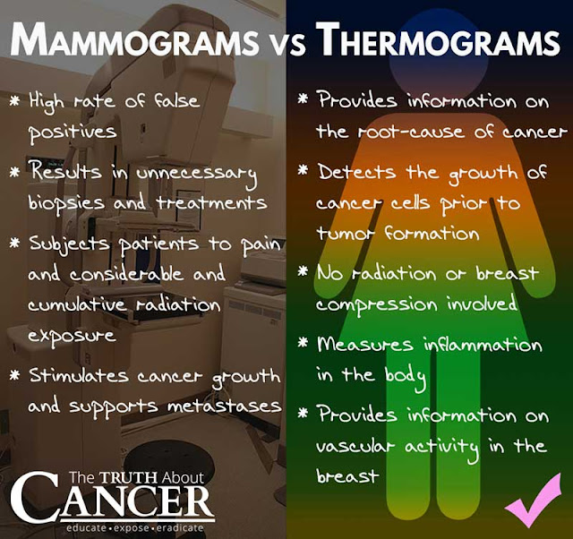
The truth is that the ionizing radiation used in mammograms are relatively high causing mutations that result in breast cancer.
One of the victims of breast cancer is the founder of BreastCancerCOnqueror.com. She is Dr. Veronique Desaulniers, a Doctor of Chiropractic. She said:
“Well, let’s start with mammograms. Again, by the time they see a lump on a mammogram, it has taken five to eight years to develop. And if a woman follows the 10-year protocol of getting a mammogram every year or every six months, she is getting as much radiation as a woman had at Hiroshima; the atomic bomb, if she stood a mile away from the epicenter. She is getting over 5 rads of radiation in a mammogram if she follows the traditional methods of mammography.”
“So, mammograms cause a lot of problems – the compression, the radiation. Studies have shown that they can increase your risk of cancer. And the new study that came out in Canada, it was a 25-year study where they found that mammograms did not decrease breast mortality rate at all. In fact, they were just as effective as a self-breast exam that women do.”
“So, if we put mammograms aside and we look at the technology with thermography, which has been around for years and can detect physiological changes at a very early stage, wouldn’t you rather know that there is possibly some changes going on in your breast or in your abdomen five to eight years before you have a diagnosis? And with thermography, there is no pain, there is no compression, there is no radiation. It is just a beautiful tool to have in your pocket.”
Thermography For Breast Cancer Screening – Accurate And Early Diagnosis
Thermography is a non-invasive and painless test, with no radiation involved. It is a test that detects and records changes in temperature on the surface of the skin.
Digital infrared thermal imaging (DITI), is a type of thermography that is used in the screening of breast cancer. It involves the use of an infrared thermal camera that takes a picture of the areas of different temperature in the breasts. The camera displays these patterns as a sort of heat map.
The presence of cancerous growth is associated with the excessive formation of blood vessels and inflammation in the breast tissue. These show up on the infrared image as areas with higher skin temperature.
How Does Thermography Work To Detect Early Cancer?
The way thermography, or thermal imaging, works is based on how cancer cells grow. When cancer cells are growing and multiplying in a tumor, blood flow is very fast in that area, blood vessels are opened, inactive blood vessels are activated and new ones are created.
Increased blood flow makes skin temperature increase and also the newly formed and activated blood vessels have a different appearance. This rise in skin temperature, as well as the presence of distinct blood vessels, is what breast thermography is aiming to detect.
Compared with other procedures (mammography) being used on their own, thermography makes it possible for cancer or pre-cancerous growth to be detected up to 10 years sooner than they may otherwise have been.
Inflammation, which is always present in precancerous and cancerous cells, also emits heat that shows up in the thermogram. That’s why, aside from cancer and inflammation, this type of screening method is also used to detect other changes in the body such as infections, allergies, trauma, fever, or flu.
Advantages Of Opting For Breast Thermography Screening
It’s Safe, And Radiation-Free
Thermography does not emit radiation – making it a safer option even for pregnant and nursing women. It just takes an image. That’s why it’s great to use as routine screening.
It’s Non-Invasive And No-Touch
There is no pressure involved that will flatten your breast or other body parts to get the imaging done. There is no pain involved. Found in the room is just an infrared camera that takes photos of your body.
It’s Very Accurate
Thermography is incredibly accurate when used as a routine screening tool.
In an article published in dontmesswithmama.com, The Institute for the Advancement of Medical Thermography reported that:
“It has been shown to be effective in finding early signs of breast cancer up to 8 years before the mammogram. As a routine screening tool, it has been shown to be 97% effective at detecting benign vs malignant breast abnormalities. Another study tracked 1,537 women with abnormal thermograms for 12 years. They had normal mammograms and physical exams. Within 5 years, 40% of the women developed malignancies. The researchers commented that an abnormal thermogram is the single most important marker of high risk for the future development of breast cancer.”
Mammograms were found to have a 65% false positive rate – which can lead to emotional stress and anxiety for patients.
It’s Key For Early Detection
Philip Getson, D.O., a medical thermographer since 1982.3 says that:
“Since thermal imaging detects changes at the cellular level, studies suggest that this test can detect activity 8 to 10 years before any other test. This makes it unique in that it affords us the opportunity to view changes before the actual formation of the tumor. Studies have shown that by the time a tumor has grown to sufficient size to be detectable by physical examination or mammography, it has in fact been growing for about seven years achieving more than 25 doublings of the malignant cell colony. At 90 days there are two cells, at one year there are 16 cells, and at five years there are 1,048,576 cells–an amount that is still undetectable by a mammogram.”
Through early detection, you can actually make changes to your diet and lifestyle before the condition develops.
Suitable For Younger Women
Premenopausal women whose breasts tend to be denser should take thermography as their first choice. This screening method can differentiate fibrocystic tissue, scars or breast implants from that of cancerous cells.
It Can Be Used For The Whole Body
Thermography can be used for:
Breast health
Deep vein thrombosis
Dental pathologies
Diabetes
Digestion and colon function
General toxicity
Immune and lymphatic system function
Inflammation
Joint health
Lung and heart health
Nerve health
Pain
Sinus infections
Thyroid health
Visceral health (liver, pancreas, spleen, and stomach)
Thermography Over Mammography For Breast Cancer Screening
A number of studies are favoring thermography for breast cancer screening for early detection, thereby making it a promising technique in the future.
There were studies showing that their experimental results on a large dataset of thermograms confirm the efficacy of the approach providing a classification accuracy of about 80%, thus taking an edge over mammography.
Furthermore, in a study reported by the juicingforhealth.com, 1,245 women were diagnosed at initial examination as either normal or benign disease by conventional means, including physical examination, mammography, ultrasonography, and fine needle aspiration or biopsy. Within five years, more than a third of the group had histologically confirmed cancers. With the help of thermography, they were was able to detect metabolic changes in the lesion cells that had the potential to grow rapidly.
Research shows that mammography gives a high probability of “false positive results.” Experts found that mammography stood for cumulative risk of 49.1 percent of false positive results on an estimate.
Meanwhile, in a study reported at the ncbi.nlm.nih.gov which compare the effectiveness of mammography, clinical examination, and thermography results showed that the greatest effectiveness of mammography vs. clinical examination was seen in the detection of early breast cancers (small lesions with negative axillary lymph nodes). In this group, thermography was less effective than it was in patients with larger lesions and lymph node metastases.
Final Words
What do you think about thermography? Well, when it comes to your health, you should always choose what’s safe and best.
Thermography, as compared to mammography, has been proven to have the potential to detect breast cancer earlier, thereby allowing you to change your lifestyle, forget your unhealthy habits, and improve your health.
It’s up to you whether you are pro-conventional, invasive method or you want the traditional, holistic, and natural approach. It’s your choice.


